History of Refractive Surgery
A TIMELINE
The History of Vision Correction
It is important to note that although many of the techniques, lasers, and lenses we use in refractive surgery are highly advanced and modern, they are all based on sound, fundamental principles, the research of which has been studied and refined over many decades. This impressive track record provides not only an interesting historical timeline, but makes modern vision correction incredibly safe and effective.
Harold Ridley, an English ophthalmologist, invents the first intraocular lens (IOL) for the treatment of cataract. During WW2, he saw many Royal Air Force casualties who had shards of material from airplane cockpit canopies lodged in their eyes. He noticed that the acrylic plastic (PMMA) splinters did not trigger an inflammatory response as did the glass splinters. The intraocular lens he produces is made of PMMA and well-tolerated by the eye.
Jose Barraquer introduces Keratomileusis. Barraquer, a Spanish ophthalmologist who did much of his research in Colombia, dedicated his life to correcting refractive errors through modifying the shape of the cornea. Keratomileusis, derived from Greek root words meaning to shape the cornea, is found to be effective. The basis of the operation is to remove, modify, and reinsert a corneal disc into the patient’s eye. The corneal disc is frozen and sculpted with a special lathe according to computer measurements. The procedure, although it worked, was technically difficult and time-consuming.
The FDA approves Radial Keratotomy (RK). First developed by Svyatoslav Fyodorov, a Russian ophthalmologist, in 1974 for the treatment of myopia. It involves making small spoke-like incisions on the cornea, which flattens its shape to correct vision. Although effective, it is no longer performed in favor of other modern procedures.
The FDA approves the Intraocular Lens (IOL) for use in cataract surgery. Prior to the use of IOLs, cataract patients had to wear very thick eyeglasses or special contact lenses in order to see after the cataract was removed during surgery.
The FDA approves the excimer laser for use in PRK, a procedure that corrects refractive error by gently preparing the surface of the eye before using the laser to reshape the cornea.
The FDA approves the excimer laser for use in LASIK. Traditional LASIK created a flap in the cornea with a manual oscillating blade, then corrected refractive error with the excimer laser. This method is not utilized at Brusco Vision.
The FDA approves the femtosecond laser for use in LASIK. The laser is used to create the LASIK flap, followed by the excimer laser to reshape the cornea. This all-laser technique is safer and more precise than using a manual blade as in traditional LASIK.

The FDA approves the use of wavefront (guided and optimized) technology for use in LASIK. With this wavefront technology, the goal of surgery is no longer to equal the best glasses or contacts vision, but in many cases to exceed the quality of vision of glasses or contacts.
The FDA approves the STAAR Visian ICL, implantable collamer lens. This biocompatible lens is placed behind the iris and in front of the eye’s natural lens to treat myopia. For many patients, this ICL provides better optics than with laser vision correction.
The FDA approves the first presbyopia-related IOL for use in cataract surgery. It is based on the principle of apodized diffraction, which bends light to two different focal points, for both near and far vision. Refractive cataract surgery and refractive lens exchange is born.

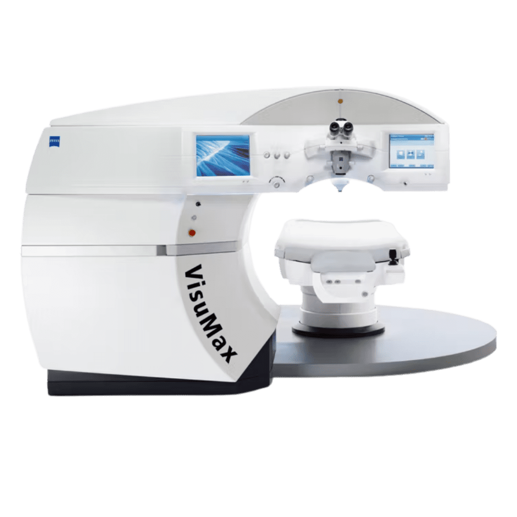
The FDA approves the Visumax femtosecond laser for use in SMILE. This procedure uses the femtosecond laser, without the use of the excimer laser, to treat myopia. It also removes less tissue than LASIK without the use of a flap.
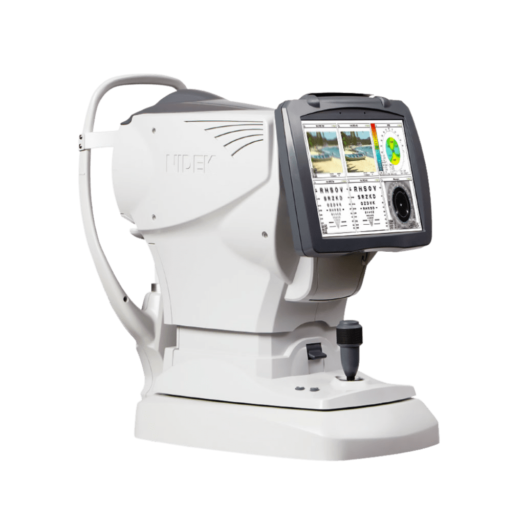
The FDA approves the use of topography-guided LASIK, which further customizes a laser vision correction procedure by providing detailed measurements mapped to each person’s corneal surface.
The FDA approves the use of the first trifocal presbyopia-related IOL for use in cataract surgery. It is based on the principle of apodized diffraction, which bends light to three different focal points - for near, far, and intermediate vision.

